Omni Cell
Imager® (OCI)
OCI-ST 8310 Standard(BF/DF/PC/DIC/Fused/Phase Imaging)
Omni Cell
Imager® (OCI)
OCI-ST 8310 Standard(BF/DF/PC/DIC/Fused/Phase Imaging)
Performance Showcase
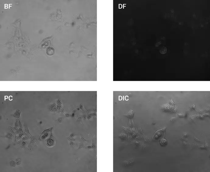
Prostate Cancer Images (BF, DF, PC, DIC)(40x)
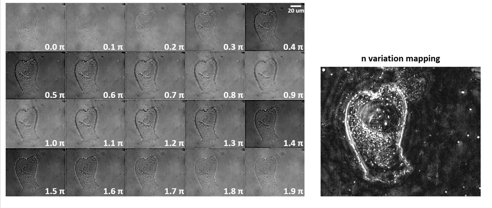
ADSC Phase Scan PC Images
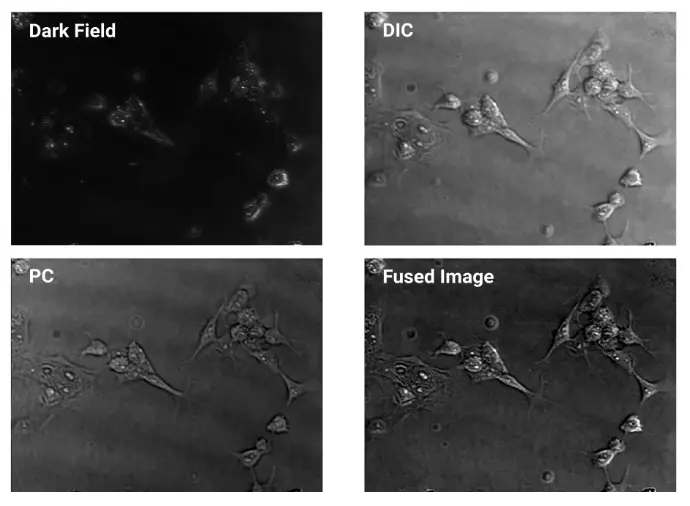
Prostate Cancer Images (DF, PC, DIC, Fused Image)
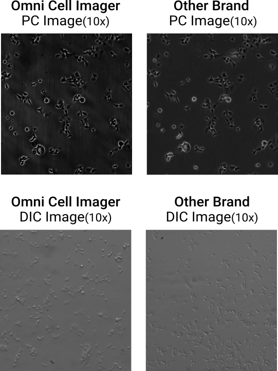
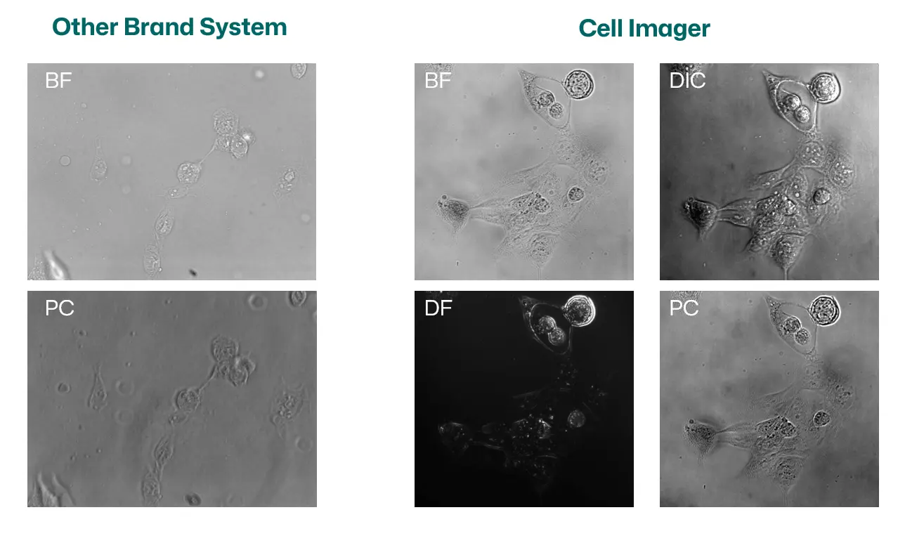
One System. Both Phase Contrast and DIC. Image Quality You Can Trust
Achieve high-contrast PC and DIC imaging on a single, software-defined microscope — with clarity rivaling traditional optical systems.
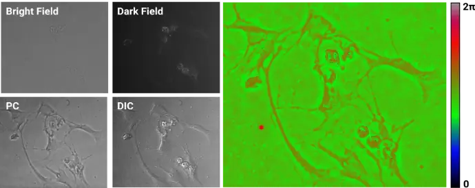
Quantitative Phase Image (QPI) with BF, DF, PC, and DIC(40x)
~0.25s to take one QPI image, ~0.6s to take all images
Prostate Cancer Images (BF, DF, PC, DIC)(40x)
ADSC Phase Scan PC Images
Prostate Cancer Images (DF, PC, DIC, Fused Image)
One System. Both Phase Contrast and DIC. Image Quality You Can Trust
Achieve high-contrast PC and DIC imaging on a single, software-defined microscope — with clarity rivaling traditional optical systems.
Quantitative Phase Image (QPI) with BF, DF, PC, and DIC(40x)
~0.25s to take one QPI image, ~0.6s to take all images

Real-Time Recording of Exosome Brownian Motion
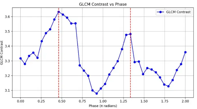
The Relationship Between GLCM Contrast and Phase
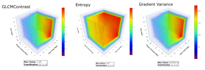
Optimal Proportion Analysis of Fused Images
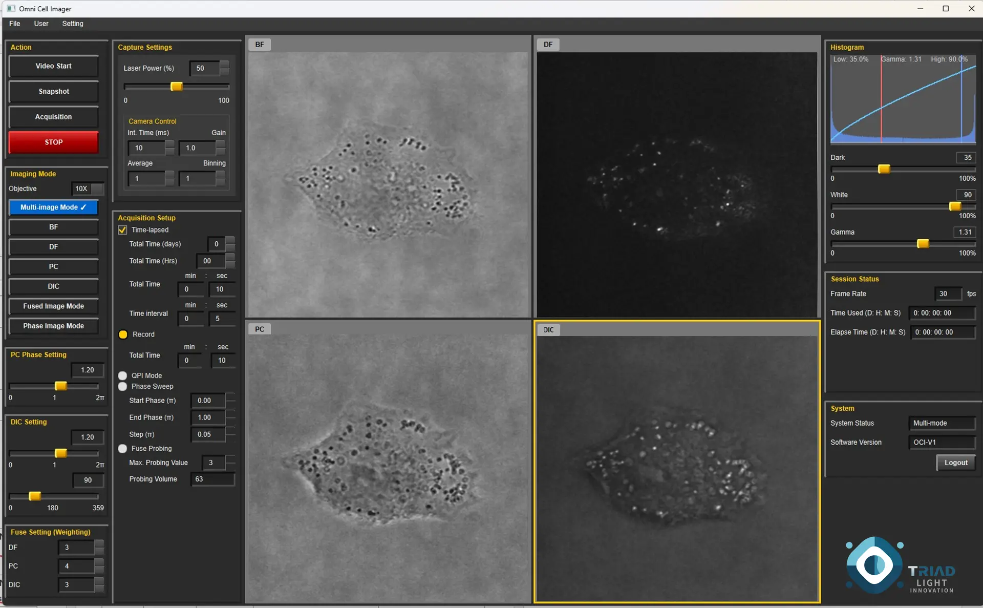
Software UI and Simultaneous Displaying 4 Modes(BF, DF, PC, DIC) of Images of The Same ROI

No More Trade-offs in Cell Imaging
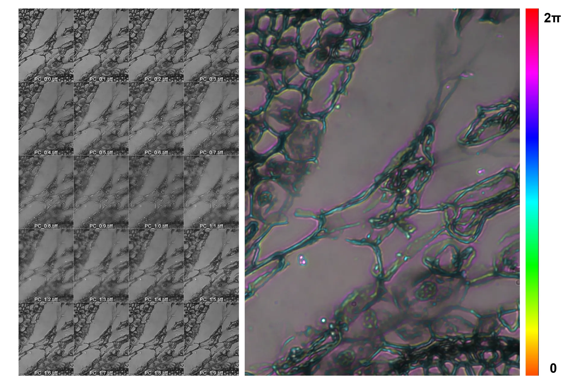
Color-coded max projection from a 0.1π phase-scan stack, capturing subtle refractive-index variations in a pine stem tissue slice
Phase Scan Phase Contrast (PSPC) Image
Real-Time Recording of Exosome Brownian Motion
The Relationship Between GLCM Contrast and Phase
Optimal Proportion Analysis of Fused Images
Software UI and Simultaneous Displaying
4 Modes of Images of
The Same ROI
No More Trade-offs in Cell Imaging
Color-coded max projection from a 0.1π phase-scan stack, capturing subtle refractive-index variations in a pine stem tissue slice